Share this Article
Doctors can see inside your arteries without cutting you open. Angiography has been quietly catching life-threatening problems before they turn catastrophic for decades.
Chest pain that comes and goes. Dizziness without explanation. Legs that ache after short walks. These symptoms send specialists toward an angiography test because what they need is not just confirmation that something is wrong but also exactly where and how bad it is.
What Is Angiography?
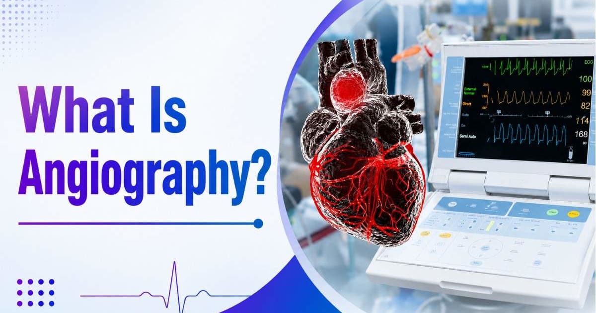
An angiography test is an imaging procedure that examines blood vessels and how blood moves through them. Standard scans cannot show vessels clearly on their own, so a contrast dye is used to make arteries and veins visible. Blockages, narrowing, leaks, and abnormal formations show up clearly once the dye is in.
Used across the heart, brain, kidneys, lungs, and limbs. Wherever blood flow is suspected to be compromised, this is usually part of the investigation.
Why Is Angiography Done?
Symptoms push the decision. When something about a patient's presentation suggests a vascular problem, this test gives doctors the specific picture they cannot get any other way.
Common reasons a doctor orders angiography:
Chest pain or suspected coronary artery disease
Investigating a potential heart blockage
Stroke evaluation or follow-up
Peripheral artery disease causing leg pain or poor circulation
Suspected aneurysm in the brain or aorta
Blood clots in the lungs or deep veins
Kidney artery narrowing affecting function
Unexplained circulation problems elsewhere in the body
The goal is always the same: find exactly what is happening and where, so treatment can be targeted and accurate.
But doctors may also recommend an ECG test before angiography to evaluate the heart's electrical activity and determine whether further investigation is needed.
Types of Angiography
Different types of angiography exist because the body has different vascular territories, and each one requires a focused approach.
Coronary Angiography
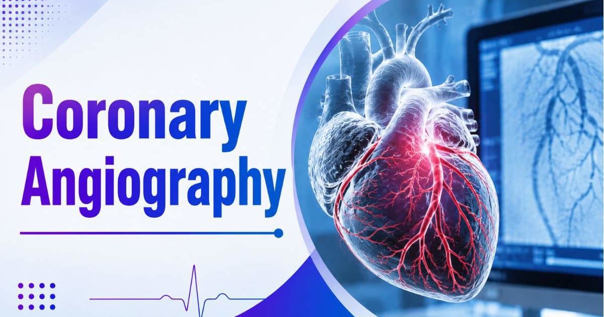
The most commonly performed type. Looks at the arteries supplying the heart muscle. Primary tool for diagnosing coronary artery disease and identifying heart blockages. Serious chest pain with elevated cardiac markers almost always leads here.
Cerebral Angiography
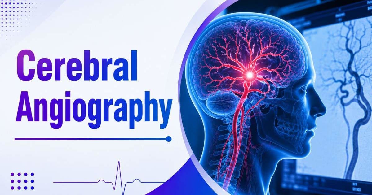
Focuses on brain blood vessels. Used for aneurysms, strokes, arteriovenous malformations, or neurological symptoms with a suspected vascular cause.
Peripheral Angiography
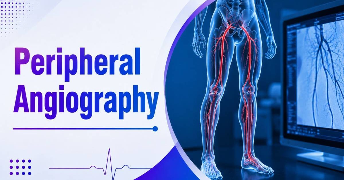
Examines arteries and veins in the limbs. Used for peripheral artery disease, non-healing wounds, or chronic limb pain to map where circulation is restricted.
Pulmonary Angiography
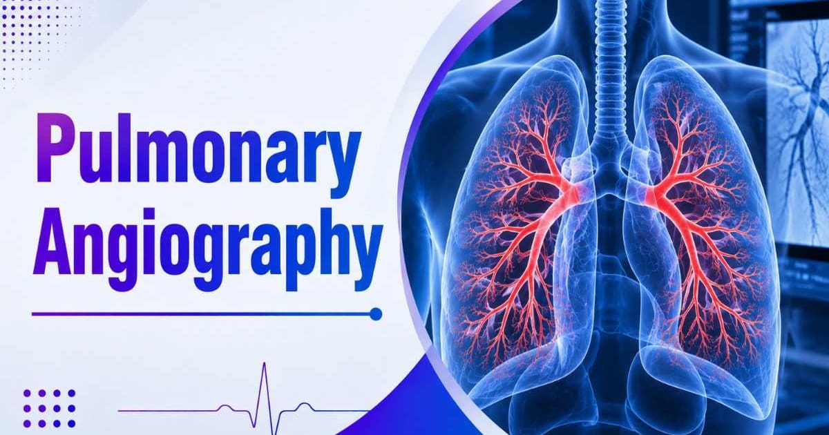
Studies the lung arteries. Historically, it was the standard for diagnosing pulmonary embolism, though CT angiography has largely replaced it in most hospitals.
Renal Angiography
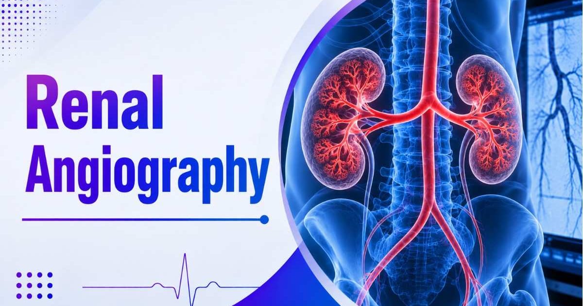
Examines the arteries feeding the kidneys. Narrowing here causes treatment-resistant high blood pressure and kidney decline that does not respond to medication normally.
Digital Subtraction Angiography (DSA)
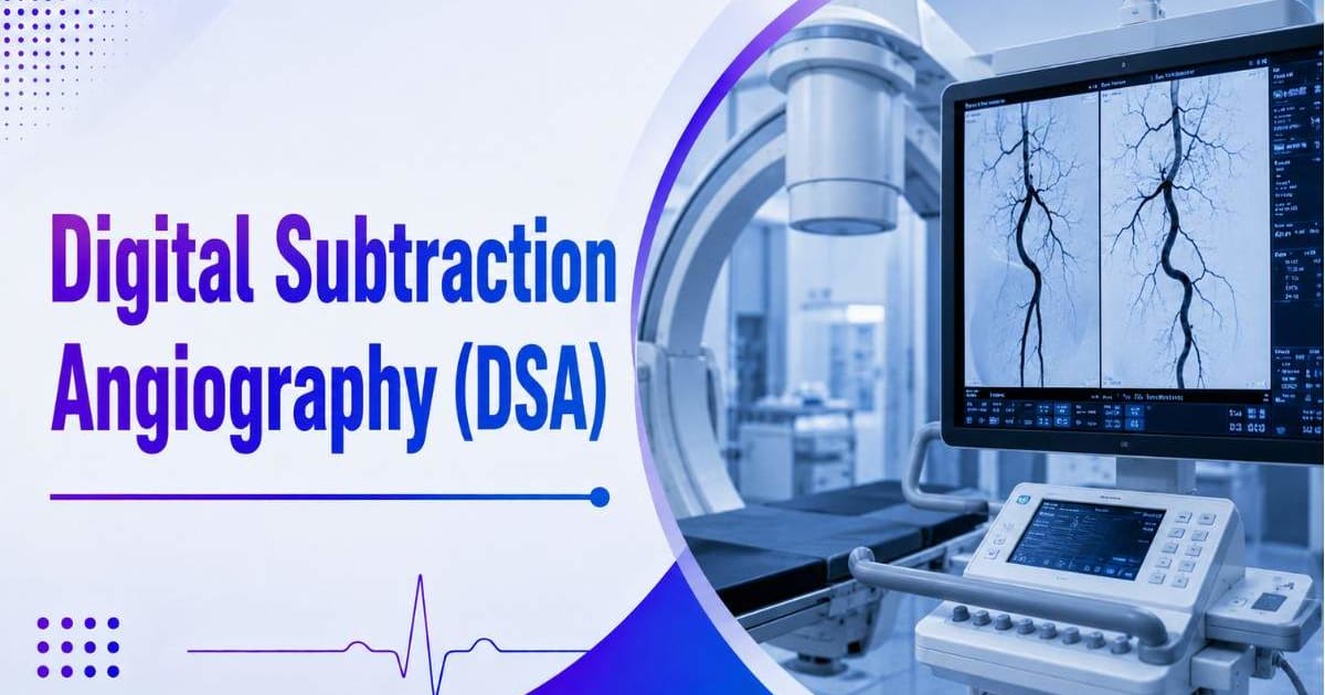
Uses software to strip bone and soft tissue from the image, leaving only vessels visible. Particularly useful for brain and peripheral studies where clarity is critical.
How does the Angiography Procedure Work?
The procedure happens in a specialised catheterisation laboratory, not a regular imaging suite.
Here is the sequence for conventional angiography:
Local anaesthesia applied to the wrist or groin where access will be made
A thin catheter inserted into the blood vessel
Catheter guided carefully toward the target area under live X-ray
Contrast dye injected through the catheter
Real-time images captured as dye travels through the vessels
Blood flow and any obstructions evaluated immediately
The whole thing takes roughly 30 to 60 minutes. Patients are awake throughout in most cases. If a significant blockage is found, the team can sometimes perform angioplasty in the same session, avoiding a second procedure.
What Is Cardiac Catheterisation?
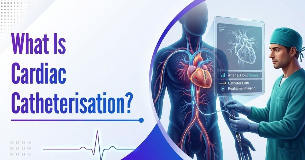
People use these terms interchangeably, but they are not the same thing.
Cardiac catheterisation is the access technique: a catheter is inserted and guided to the heart. Coronary angiography is the imaging study performed through that access. One is how you get there. The other is what you do once you arrive.
CT Angiography vs Conventional Angiography
CT Angiography | Conventional Angiography |
Non-invasive, no catheter | Minimally invasive, requires catheter |
Uses CT scanner with IV dye | Uses catheter and live X-ray imaging |
Faster, good for screening | More detailed, real-time assessment |
Cannot treat during same session | Can proceed directly to angioplasty if needed |
Lower risk profile | Considered the diagnostic gold standard |
CT angiography suits screening and situations where intervention is not expected. Conventional angiography is the standard when treatment may follow immediately or when CT detail is insufficient.
How to Prepare for an Angiography Test?
Standard preparation instructions:
Fast for six to eight hours beforehand
Tell the doctor about all current medications
Flag any known allergy to iodine or contrast dye in advance
Avoid caffeine before heart-related scans
Stay hydrated unless told otherwise
Specific instructions vary by procedure type. The treating team will advise accordingly.
Is Angiography Painful?
Local anaesthesia handles the insertion site. The initial needle prick is the most uncomfortable part. When contrast dye goes in, a brief warm flushing sensation passes within seconds. Most patients describe the procedure as tolerable. The anticipation is usually worse than the reality.
Risks and Complications
Possible complications include the following:
Minor bleeding or bruising at the catheter insertion site
Infection
Allergic reaction to the contrast dye
Temporary strain on the kidneys from the dye
Irregular heart rhythms during the procedure
In rare cases, stroke or heart attack
Serious complications are uncommon with experienced specialists in a proper facility.
Angiography Cost in India
Costs vary by procedure type, hospital, and city.
Procedure | Estimated Cost |
CT Angiography | Rs. 7,000 to Rs. 16,000 |
Conventional Angiography | Rs. 15,000 to Rs. 60,000 |
Basic Diagnostic Angiography | Rs. 6,000 to Rs. 20,000 |
Digital Subtraction Angiography | Varies by hospital and complexity |
These figures typically cover imaging charges and catheterisation lab costs. Doctor fees, consumables, and any observation period after the procedure add to the total. India draws international patients for the usual reasons: qualified specialists and costs well below Western equivalents.
What Happens After Angiography?
After conventional angiography, patients are observed for a few hours. Groin access needs a bit more rest than wrist access to reduce bleeding risk. Wrist access usually allows same-day discharge.
Post-procedure advice typically includes:
Drink plenty of fluids to help clear the contrast dye
Avoid heavy lifting for a short period
Watch the puncture site for unusual bleeding or swelling
Attend follow-up appointments as scheduled
Most people are back to normal activity within one to two days. For patients diagnosed with severe coronary artery disease, follow-up care is important to reduce the risk of complications such as heart failure.
Conclusion
Angiography remains one of the most precise diagnostic tools available when blood flow is in question. Whether it is coronary angiography identifying a heart blockage, cerebral angiography mapping brain vessels, or DSA providing a clear view of peripheral circulation, the technology continues to improve. It also helps doctors evaluate reduced blood flow in patients with conditions such as cardiomyopathy, allowing more accurate treatment planning.
Knowing what the procedure involves and what to expect afterward can make the experience far less stressful. If you're looking for trusted hospitals, experienced specialists, or guidance on angiography and other medical procedures, GetBeds can help you explore the right treatment options and make informed healthcare decisions.
Frequently Asked Questions
An imaging test that uses contrast dye to make blood vessels visible on X-ray or CT scans. Done to diagnose blockages, narrowing, aneurysms, blood clots, and other vascular problems affecting the heart, brain, kidneys, lungs, or limbs.
A catheter is inserted through the wrist or groin under local anaesthesia, guided to the target blood vessel, contrast dye is injected, and real-time X-ray images capture blood flow and any abnormalities. Takes 30 to 60 minutes in most cases.
Coronary, cerebral, peripheral, pulmonary, renal, and digital subtraction angiography. Each targets a different part of the vascular system depending on where the suspected problem is.
G
Generally safe with low complication rates when performed by experienced teams. Minimally uncomfortable for most patients. Local anaesthesia handles the insertion site, and the contrast dye causes a brief warm sensation that fades within seconds.
CT angiography runs roughly Rs. 7,000 to Rs. 16,000. Conventional angiography falls between Rs. 15,000 and Rs. 60,000. Final costs depend on procedure type, hospital, and city.
CT angiography is non-invasive and uses a scanner with intravenous dye, better suited for screening. Conventional angiography uses a catheter and provides real-time imaging considered the diagnostic gold standard, with the added option of treating a blockage in the same session if one is found.
Disclaimer:
This blog is intended for informational purposes only and does not constitute medical advice. Always consult a qualified healthcare professional for diagnosis and treatment.